MRI Scan
Shree Imaging is well known for bringing in the latest scanning techniques in Thane and Mumbai. We are amongst the few centres to perform MR Mammography, MR Tractography, Cardiac MRI.
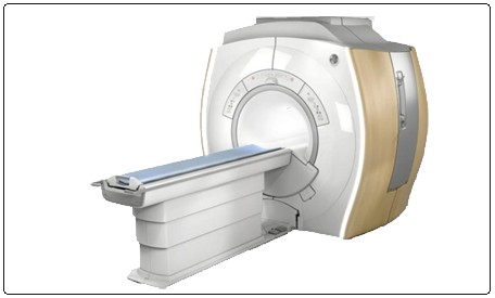
Our centre is equipped with the latest high-Definition 1.5 Tesla MRI system from GE Healthcare and all MR examinations, including ultrafast sub-second scans are routinely performed. Even uncooperative patients can be scanned using special PROPELLOR sequences
What is MRI?
MRI is a non-invasive medical imaging modality using nuclear magnetic resonance to demonstrate pathological or other physiological alterations. MR imaging uses a powerful magnet, radio waves and a computer to generate pictures of body structures in exquisite detail. MRI uses movement of hydrogen nuclei in water and fat within the body to generate images from complex mathematical data ( 2D or 3D matrix).
No harmful ionizing radiation is used while performing MRI.Precautions before entering MRI Room
You must inform the technician if you have an implant, pacemaker, artificial heart valve. The magnetic resonance will interfere with these medical devices.
The usual applications of MRI are in:
- Diffusion weighted images are very commonly used in the assessment of acute stroke and are also widely used in oncology.
- MR angiography is used to generate pictures of the arteries to detect any abnormal narrowing/dilatation or abnormal arterio-venous connections. MR angiography is often used to evaluate the arteries of the neck and brain. Magnetic Resonance Venography (MRV) is a similar procedure used to image veins.
Diffusion Tensor Imaging , also known as MR Tractography non-invasively maps white matter tracts. a powerful tool to study brain tumors, Alzheimer's disease and many more.
- MR Angiography is used to evaluate thoracic and abdominal vessels, renal arteries and lower limb arteries.
- MRCP is used to evaluate the hepato-biliary system and pancreas.
- MR fistulography and defecography is technique to study disorders of ano-rectal function and infections. It is indicated in patients having constipation, rectal incontinence, painful defection and rectal prolapse. Conventional studies do not provide the complete three dimensional anatomical information.
- MR imaging provides information regarding the ligaments and tendons along with the muscles in and around the joints.
Visualizing degenerative disease of spine and bulging discs.
- Although X-Ray Mammography remains the primary imaging modality in the evaluation of breast disease, the mammogram can at times be inconclusive.
- MR imaging has been used as an adjunct to mammography, particularly for patients with equivocal mammographic findings. The main advantage of breast MR imaging is its high sensitivity, with reported sensitivities for cancer ranging from 91% to 100% with varying specificity.
- It can be useful to verify multicentricity and multifocality of breast cancer, differentiate scars from recurrences after breast-conserving therapy, screen high-risk groups, investigate breast implants.
- Common cardiac pathologies evaluated on MRI are usually ischemic heart disease, myocarditis, cardiomyopathies, valvular heart disease, congenital heart disease and cardiac tumors.